How a gut-bacterial protein makes β-cells grow
My Ph.D. work in the Guillemin Lab asks how microbes shape pancreatic β-cell development, and whether one bacterial protein could help expand or mature β-cells for diabetes therapy. NIH NIDDK F31 fellowship, 2025–2027.
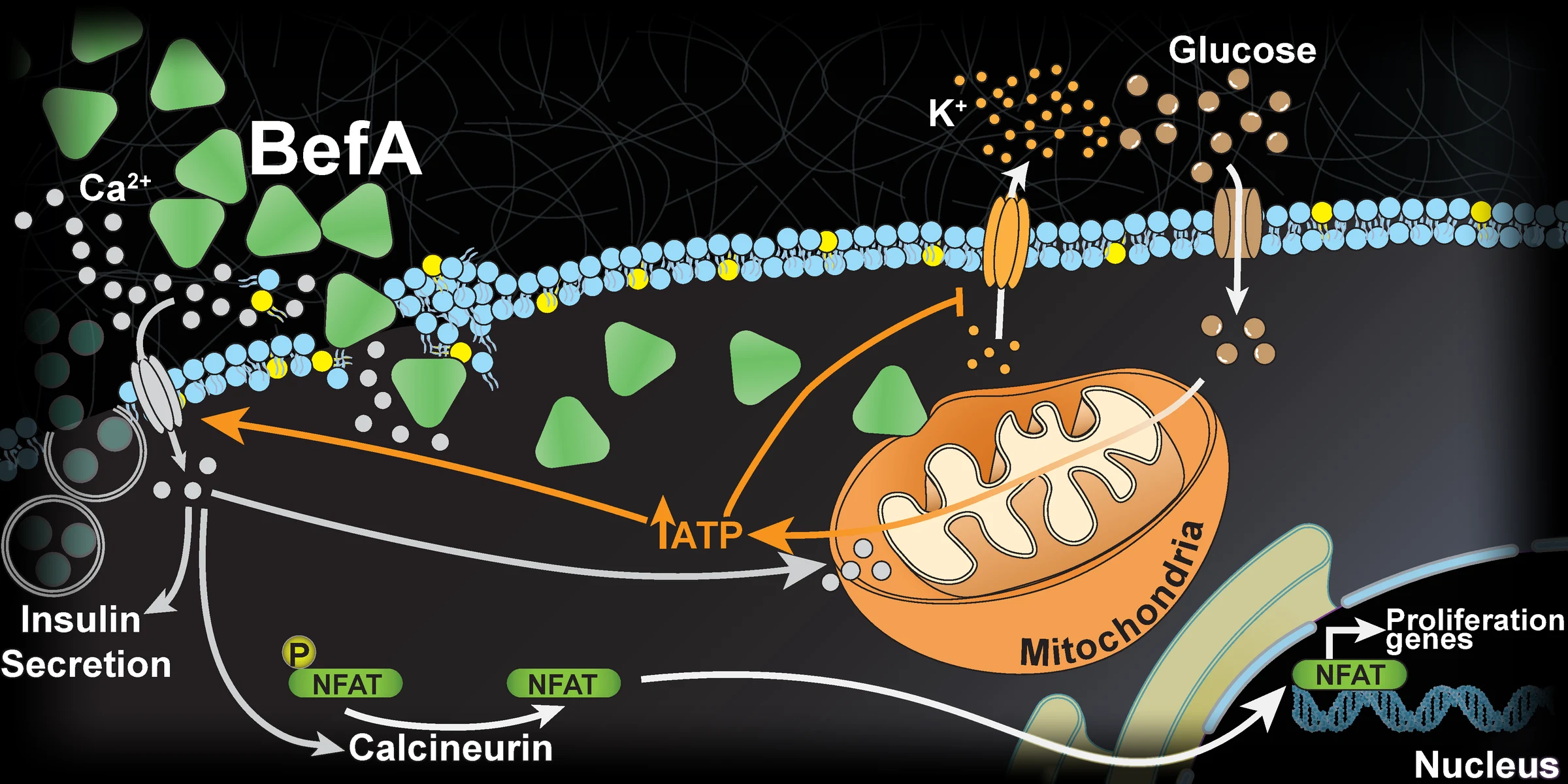
- Membrane
BefA permeabilizes the β-cell membrane.
- Calcium
Calcium floods into the cell.
- Mitochondria
Mitochondrial function and ATP output rise.
- Proliferation
Calcineurin–NFAT signaling drives β-cell proliferation.
The question
β-cells make insulin. Lose them and you get type 1 diabetes; wear them out and you get type 2.
Yet we still understand little about what makes β-cells grow and mature early in life. Gut microbes turn out to matter: germ-free zebrafish develop fewer β-cells, and a single secreted bacterial protein, BefA (Beta-cell Expansion Factor A), is enough to restore them (Hill et al., 2016; Hill et al., 2022).
My dissertation asks how BefA does it, at the level of the membrane, the cell, and the whole animal.
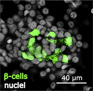 Germ-free
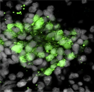 BefA
What I found
BefA acts directly on β-cells.
Combining genetic and pharmacological manipulation, live super-resolution imaging, single-cell RNA-seq, and mitochondrial respirometry, in zebrafish, ex vivo neonatal mouse islets, and cultured mammalian cells, I found that BefA permeabilizes β-cell membranes, drives calcium flux, and promotes mitochondrial function. The result is β-cell proliferation and improved metabolic potential.
- Live super-resolution & TIRF imaging
- scRNA-seq
- Mitochondrial respirometry
- Zebrafish genetics
- Ex vivo mouse islets
- Cultured β-cells
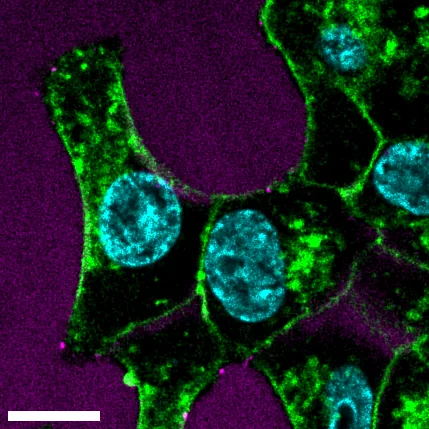 mScarlet
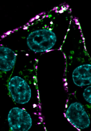 mScarlet-BefA
Where it lands
β-cell proliferation can be induced with an exogenous protein.
That points to two applications: prophylactically expanding β-cells early in life to help prevent diabetes, and maturing induced pluripotent stem cell (iPSC)-derived β-cells for transplant.
The work also supported two funded NIH grants for the lab: an R01 on BefA's mechanism and therapeutic potential ($1.29M), and an R61 on engineering mitohormetic probiotics ($1.03M).
Before the Ph.D.
Environmental pathogen surveillance
Led the Thermo Fisher partnership that took a prototype air sampler through five Design-of-Experiments validation phases to commercial release as the AerosolSense sampler, extended it to influenza A, influenza B and RSV, and showed that building air can serve as a real-time, non-invasive readout of infection indoors.
Zebrafish models of patient variants
Built CRISPR/Cas9 knockout and knock-in zebrafish carrying the exact alleles of families in the NIH Undiagnosed Diseases Network, giving clinicians functional evidence on variants of uncertain significance.